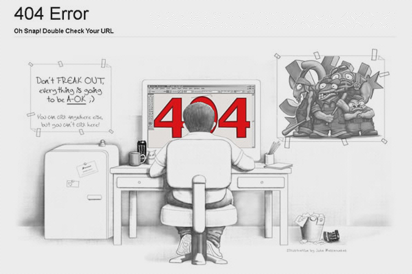
特发性脊柱侧弯畸形的分型与治疗.pptx

上传人:h**** 文档编号:230969 上传时间:2018-07-25 格式:PPTX 页数:35 大小:2.30MB
下载 相关 举报
特发性脊柱侧弯畸形的分型与治疗.pptx_第1页
第1页 / 共35页
特发性脊柱侧弯畸形的分型与治疗.pptx_第2页
第2页 / 共35页
特发性脊柱侧弯畸形的分型与治疗.pptx_第3页
第3页 / 共35页
特发性脊柱侧弯畸形的分型与治疗.pptx_第4页
第4页 / 共35页
特发性脊柱侧弯畸形的分型与治疗.pptx_第5页
第5页 / 共35页
点击查看更多>>
资源描述

1、Adolescent Scoliosis Classification and Treatment特发性脊柱侧弯畸形的分型与治疗,Jane S. Hoashi, MD, MPH, Patrick J. Cahill, MD,James T. Bennett, MD, Amer F. Samdani, MD*,Neurosurg Clin N Am 24 (2013) 173183,KEYWORDS,Adolescent idiopathic scoliosis Lenke classification Scoliosis Pediatric spine deformity Pedicle sc

2、rews青少年特发性脊柱侧弯Lenke分型弓根螺钉矫形,KEY POINTS,Adolescent idiopathic scoliosis (AIS) can be classified according to the Lenke classification system, which incorporates curve magnitude, flexibility, the lumbar modifier, and the sagittal plane.青少年特发性脊柱侧凸(AIS)可根据Lenke分类系统进行分类,该系统包括曲线大小,柔韧性。The Lenke classifica

3、tion serves as a guide with respect to level selection in patients with AIS.Lenke分类可作为AIS患者融合水平选择的指南。The widespread use of pedicle screws has resulted in most AIS being treated through a posterior approach.椎弓根螺钉的广泛使大多数AIS可以用后路治疗。,INTRODUCTION,Adolescent idiopathic scoliosis (AIS) is a spinal conditi

4、on causing deformity of the spine in 3 dimensions: the coronal, sagittal, and axial planes. AIS is defined as any curve equal to or greater than 10 in the coronal plane1,2 in patients 10 to 18 years old.3 It is a diagnosis of exclusion after congenital, neuromuscular, neural, or syndromic causes of

5、scoliosis have been ruled out. Preoperative mag-netic resonance imaging is useful for ruling out neural causes of scoliosis, such as syringomyelia or Chiari malformation, although its use as a preop-erative screening tool is controversial.4,5 A genetic component has been described regarding the caus

6、e of AIS.611 With an incidence of 11% among first-degree relatives,12 it is not uncommon for a health care provider to manage multiple mem-bers of a family with scoliosis.青少年特发性脊柱侧凸(AIS)是一种脊柱疾病,在三维方面引起脊柱畸形:冠状面,矢状面和轴面。 AIS被定义为10-18岁患者冠状面等于或大于10。排除先天性,神经肌肉,神经或综合征引起的脊柱侧凸原因。 术前磁共振成像对于排除脊柱侧凸的神经原因,如脊髓空洞症或

7、Chiari畸形是有用的,尽管其作为术前筛查工具的使用还存在争议.已经报道了AIS的原因 .在一级亲属中,发生率为11,医疗保健提供者报道一个家庭有多个脊柱侧弯患者的情况并不少见。,AIS affects approximately 2% to 3% of the adolescent population, but fewer than 10% of patients with AIS need treatment.13 The higher the curve magnitude, the lower the prevalence and the higher the female/mal

8、e ratio. Curves greater than 30 have a 0.1% to 0.3% prevalence and affect females 10 times more than males.AIS对青春期人群的影响约为23,而AIS患者中只有不到10需要治疗。曲度越重,患病率越低,女性比例越高。曲度大于30的患病率为0.10.3,女性患病率是男性的10倍以上。,For years, the King-Moe classification was the most widely used system for guiding treatment in AIS. Its s

9、hortcomings included classifying curves based only on the coronal plane and showing low interobserver reliability.15 Also, only variants of the thoracic curve were described, leaving some other curve types such as thoracolumbar or lumbar curves unable to be classified by this system. The Lenke class

10、ification16 addresses these shortcomings and is now considered the gold standard for classifying AIS and guiding treatment. In this article, the Lenke classification is used to describe the AIS types and the treat-ment options.多年来,King-Moe分类是用于指导AIS治疗的最广泛使用的系统。 其缺点是包括仅仅根据冠状面分型,并显示出较低的观察者间的可靠性。另外,仅描述

11、了胸弯的变体,残留了一些其他曲线类型,如胸腰弯或腰椎弯无法通过该系统进行分类。Lenke分类解决了这些缺点,现在认为是AIS分类和指导治疗的金标准。在本文中,Lenke分类用于AIS类型和治疗选择。,Treatment of scoliosis includes nonoperative management such as bracing of curves measuring 20 to 40 or progressing more than 5 per year. Larger curve magnitude, younger chronologic age, and Risser si

12、gn are associated with curve progression.17 The literature has shown bracing to be more effective in patients with earlier Risser scores (01) and open triradiate cartilages.1820 The goal of bracing is to maintain curve magnitude throughout a patients growth period, although conflicting evidence of i

13、ts effectiveness have been reported.治疗脊柱侧弯包括非手术治疗:20至40度的曲度或每年5度以上的曲度进展。 较大的曲度,较小的年龄和Risser征与曲度进展有关。文献显示早期Risser评分(0-1)和开放性Y软骨患者的支具更有效。支具的目标是保持患者在整个生长期中保持目前曲度的幅度,尽管目前的报道对其有效性的报道是相互矛盾的。,Surgery is indicated when a curve is progressive despite bracing and generally when the curve rea-ches 45 to 50 . T

14、he main goal is to stop the curve from progressing, leading to potentially severe complications from an untreated curve, including pulmonary function and back pain. Other goals driven by the patients themselves are improvement of cosmesis. Quality of life studies as measured by the SRS-22 (Scoliosis

15、 Research Society 22) ques-tionnaire have shown that patients with AIS have lower self-image and are more self-conscious about their general appearance than the general population.21,22 This finding can be related to a shoulder imbalance, rib prominence, or trunk asymmetry. Thus, the psychological i

16、mpact of the deformity must also be taken into account when considering surgery.尽管有支具,曲度仍然是进行性发展的,通常曲度大于45到50之间时表示需要手术。 手术的主要目标是阻止曲度继续进展,导致包括肺功能和背部疼痛在内的潜在的严重并发症。 患者自己的目标是改善外观。根据SRS-22的调查问卷所测量的生活质量研究显示,AIS患者的自我形象评价较低. 可能与肩部不平衡,肋骨突出或躯干不对称有关。 因此,在考虑手术时也必须考虑到畸形的心理影响。,The goals of surgery are to restore

17、coronal and sagittal balance, reduce the rib prominence, and achieve shoulder balance. However, another important goal is to leave as many unfused seg-ments as possible to preserve motion in the lumbar spine. The specific treatment options are discussed further in this article.手术的目标是恢复冠状和矢状平衡,减少肋骨突出

18、,达到肩部平衡。 然而,另一个重要的目标是尽可能多的保留未融合的部分以保持腰椎运动。 本文将进一步讨论具体的处理措施。,Two approaches to AIS surgery exist: the anterior approach and the posterior approach; a combina-tion of the 2 is also used. Some potential advan-tages to the anterior approach are saving fusion levels,23,24 decreased prominence of instrume

19、nta-tion, and decreased risk of crankshaft phenom-enon in a skeletally immature adolescent.16,25 However, some studies have indicated morbidity related to decreased pulmonary function,26,27 which seems to improve at 2-year follow-up.28 The anterior approach can be used to fuse simple thoracic curves

20、 and can also be used to perform anterior release and fusion combined with posterior spinal fusion in stiffer and larger (90 ) curves, although similar curve correction can be achieved in these larger curves by the posterior approach alone.AIS手术有两种方法:前路手术和后路手术;两种手术的组合也被使用。 前路手术的一些潜在优势是节约融合水平,降低青少年骨骼

21、不成熟的曲轴现象的风险。然而,一些研究表明发病率与 肺功能下降26,27,在2年的随访中似乎有所改善。前路手术可用于融合简单的胸弯,也可用于前路松解后路脊柱融合。,Since the development of pedicle screws, the posterior-only approach has become the mainstay of treatment of AIS. Pedicle screws provide a 3-column fixation that permits greater curve correction and improved derotation

22、.30 Even in the more severe (90 ) and stiffer curves, pedicle screw constructs with osteotomies render good correction,29 thereby reducing the need for combined anterior and posterior approaches. The crankshaft phenomenon may also be reduced by using pedicle screws.自从椎弓根螺钉发展以来,后路手术已成为AIS治疗的主要手段。 即使在

23、严重的( 90)和僵硬的侧弯治疗中,用截骨加椎弓根螺钉能得到良好的效果,从而减少对前后联合手术的依赖。 曲轴现象也可以通过使用椎弓根螺钉减少。,However, pedicle screw placement has a learning curve, especially with the free hand technique.32 With surgeon experience, the accuracy of pedicle screw placement improves, and the medial breach rate decreases.33,34 Reported bre

24、ach rates range from 1.6% to as high as 58%.3338 However, rates for neurologic and visceral injuries despite these breaches are low. Although hypokyphosis has been observed with posterior-only pedicle screw constructs,39,40 long-term follow-up has shown good maintenance of correction and coronal and

25、 sagittal alignment.然而,椎弓根螺钉置钉需要有学习曲线特别是徒手置钉技术。随着外科医生的经验提高,椎弓根螺钉置入的准确性提高,内侧破口率降低。报告的破口率从1.6到58。神经和内脏损伤的发生率很低。 只有后路椎弓根螺钉矫形才会出现交界后凸,但长期随访显示良好的矫正和冠状位及矢状位序列。,LENKE CLASSIFICATIONOverview,The Lenke classification for AIS was developed as a tool to help surgeons classify curve types and guide them in oper

26、ative treatment.16 The curve type (the major curve), lumbar modifier (A, B, and C, depending on the location of the center sacral vertical line CSVL in relation to the apical lumbar vertebra), and the sagittal profile (, N, 1) is used to determine a specific curve pattern. Although there are 6 Lenke

27、 curve types, a total of 42 curve patterns can be observed.对于AIS的Lenke分型是为了帮助外科医生对侧弯的曲线类型分类并指导他们进行手术治疗而开发的.侧弯类型(主弯),腰椎修正型(A,B和C,CSVL相对于腰椎顶椎的位置)和后凸( - ,N,1)用于确定特定的侧弯模式。 虽然有6个Lenke主弯类型,但总共可以观察到42个侧弯模式。,The basis of surgical treatment is to fuse only the structural curves. The curve with the largest Co

28、bb magnitude is defined as the major curve, which, by definition, is structural. Curves with lesser magni-tude (minor curves) can be structural or nonstruc-tural, depending on the degree of their flexibility seen on bending films. Generally, minor curves are not considered part of the arthrodesis if

29、 they bend out to less than 25 . Focal kyphosis is also a criterion for considering a curve to be structural.手术治疗的基础是只融合结构弯。 COBB最大的弯曲被定义为主弯,根据定义它是结构性的。 曲度较小的弯曲(次弯)可以是结构性的或非结构性的,这取决于它们在 bending 上看到的柔韧程度。 一般来说,如果 bending 小于25,次弯不融合。 后凸也是考虑曲线结构的标准。,The Lenke classification differentiates King-Moe type

30、 2 curves into Lenke types 1 and 3, helping surgeons select which curves are amenable to selective fusions (Lenke type 1) and those that require an extended fusion in the lumbar spine (Lenke type 3). Unlike the King-Moe classification, which considers only the coronal plane, the Lenke classification

31、 accounts for both coronal and sag-ittal planes and has been shown to have good interobserver reliability. However, the axial plane (a reflection of vertebral body rotation) is still not included in the Lenke classification. Moreover, some curve types such as curves with C lumbar modifiers are subje

32、ct to controversy regarding selective versus nonselective fusion. The following section on the specific Lenke curve types includes some of the controversies and current recommen-dations for treatment.Lenke分类将King-Moe 2型曲线区分为Lenke 1型和3型,帮助外科医生选择适合选择性融合(Lenke 1型)和需要在腰椎(Lenke 3型)中进行融合。 与仅考虑冠状面的King-Moe

33、分类不同,Lenke分类既包括冠状平面也包括矢状平面,并且已被证明具有良好的观察者间可靠性。 然而,Lenke分类仍不包括轴面(椎体旋转的反映)。 此外,某些曲线类型(如带有腰弯修正型的曲线)在选择性与非选择性融合方面存在争议。 以下关于特定Lenke曲线类型的部分包括一些争议和当前的治疗建议。,Treatment of Lenke Curve TypesLenke 1: single thoracic curve,For single thoracic curves (Fig. 1), it is generally accepted to perform selective fusions

34、 of the main thoracic curve, unless there is a kyphosis of more than 20 in the thoracolumbar area, in which case, the lumbar curve is also included in the fusion.16 The unfused lumbar curve is nonstruc-tural and usually spontaneously corrects itself after thoracic fusion.4246 It is important to note

35、 any preoperative shoulder height discrepancy, be-cause this often determines the upper fusion levels. Shoulder height can be determined clini-cally as well as radiographically using the clavicle angle or T1 tilt.对于单胸弯(图1),一般认为胸弯选择性融合是可行的,除非在胸腰段有超过20的后凸畸形,这种情况下,腰弯也需要融合16。腰椎不融合,通常在胸椎融合术后自行矫正。重要的是要注意术

36、前肩高的差异,因为这通常决定了融合的高度。 可以临床确定肩高,也可以使用锁骨角或T1倾斜进行放射学检查。,Three different scenarios exist regarding shoulder height. The first and most common scenario is a right main thoracic curve, with the right shoulder being higher than the left. In this case, correction of the thoracic spine also brings down the r

37、ight shoulder, usually achieving equal shoulder height. In these cases, the upper instru-mented level is usually T4 or T5.48 If the left shoulder is elevated, the compensatory proximal thoracic curve is usually included in the fusion (to T2) to oppose the corrective forces being placed on the main t

38、horacic curve, which would otherwise continue to drive the left shoulder up. If both shoul-ders are equal in height preoperatively, T3 is usually the upper level of fusion.关于肩高有三种不同的情况。 第一种也是最常见的情况是右侧主胸弯,右肩高于左侧。 在这种情况下,矫正胸弯也会使右肩下垂,通常达到肩高相等。 在这些情况下,UIV通常为T4或T5.如果左肩高,则补偿性近端胸椎融合通常融合T2,否则会继续向左。 如果术前双方肩高

39、相等,T3通常是UIV。,For single thoracic curves with minor flexible lumbar curves (Lenke 1A and 1B), selective thoracic fusions are generally indicated. For distal fusion levels, it is important to choose the appropriate lowest instrumented vertebra (LIV) so as to leave good coronal balance and avoid lumbar

40、 decom-pensation or progression of the primary curve (adding-on). Conventional guidelines have used the stable vertebra, or the most proximal vertebra with pedicles most closely bisected by the CSVL as the LIV.15 However, this guideline was based on Harrington instrumentation, in which the correc-ti

41、ve forces were uniplanar. With 3-column fixation using pedicle screws, an additional 1 or 2 distal motion segments can be saved, instead of fusing to the stable vertebra.对于具有较小腰弯的单胸弯(Lenke 1A和1B),一般选择性胸椎融合。 对于远端融合水平,重要的是选择合适的LIV,以保持良好的冠状平衡并避免腰椎退化或附加现象。 常规的指南使用了稳定椎。然而,这个指南是基于Harrington,其矫正力是单平面的。 通过使

42、用椎弓根螺钉可以进行三柱固定,可以节省额外的1或2个远端运动节段,而不是融合到稳定的椎骨上。,Adding-on(附加现象),2000年由Suk最先报道发生率:2-21%再次手术率为 7.3%,Adding-on(附加现象),定义:末次随访时主弯的LEV向LIV远端移动并且冠状面 Cobb角增加5;LIV远端邻近椎间盘成角增加5;LIV偏离CSVL增加10mm以上。,The neutral vertebra is also used to determine the distal fusion level.49,50 The relation between the neutral verte

43、bra and the end vertebra can be used to ascertain the LIV. If there is no more than 1 level between the end vertebra and the neutral vertebra, then fusion to the neutral vertebra is suffi-cient. This level corresponds to 1 level proximal to the stable vertebra. However, if the neutral vertebra is 2

44、or more levels distal to the end vertebra, then the LIV is NV-1. If the neutral vertebra is the end vertebra, then it is adequate to fuse to the distal end vertebra. A 2-year follow-up by Suk and colleagues49 in patients treated using these guide-lines showed satisfactory results with good coronal b

45、alance, compensatory lumbar straightening, and no adding-on.中立椎也用于确定远端融合。中立椎和端椎之间的关系可以用来确定LIV。 如果端椎和中立椎之间的距离不超过1个椎体,则融合到中立椎是足够的。 当术前NV与EV距离为两个椎体以上时,LIV选择在NV-1。 如果中性椎骨是端椎骨,则足以融合到端椎。 Suk及其同事对使用这些指南治疗的患者进行为期2年的随访,结果令人满意,具有良好的冠状平衡,腰椎矫正,无附加功能。,With regard to adding-on, Miyanji and colleagues51 differenti

46、ated 2 types of Lenke 1 curves, depending on the L4 tilt: 1A-L (tilted to the left) and 1A-R (tilted to the right). 1A-R curves have been shown to have a higher risk of adding-on because of the overhanging curve pattern, requiring a more distal fusion, approximately 2 levels more distal than a 1A-L

47、curve.关于附加现象,Miyanji和他的同事根据L4倾斜:1A-L(向左倾斜)和1A-R(向右倾斜)区分了2种类型的Lenke 1曲线。 已经显示1A-R曲线具有较高的附加风险,需要更远端的融合,比1A-L曲线更远2个节段。,Lenke 1C curves have been subject to ongoing controversy regarding their fusion levels because often they behave like double major curves. In the 1C pattern, the nonstructural lumbar c

48、urve is flexible (side-bending to 25 ), in which the apex completely crosses the midline. A study by Lenke and colleagues53 showed that selective thoracic fusion was performed in 62% of patients with 1C curves, implying that the remaining 38% had nonselective fusions. Newton and colleagues reported

49、that larger preoperative lumbar curve magnitude, greater lumbar apical vertebra dis-placement from the CSVL, and smaller thoracic/ lumbar magnitude ratio were factors associated with nonselective fusion. Lenke and colleagues55 reported that for a selective fusion to be success-ful for 1B and 1C curv

50、es, the thoracic/lumbar ratios for Cobb magnitude, apical vertebral trans-lation, and apical vertebral rotation should be greater than 1.2。Lenke 1C曲线因其融合程度而受到持续的争议,因为它们通常表现为两个大弯。 在1C模式中,非结构性腰部曲线是柔性的(side-bending25),其中顶点完全穿过中线。 Lenke等的一项研究显示62的1C曲线患者进行了选择性胸段融合,这意味着剩下的38是非选择性融合。 Newton及其同事报道,较大的术前腰弯曲度,较大的腰椎顶椎椎体位移与较小的胸椎/腰椎大小比例是非选择性融合的相关因素。 Lenke等报道,对于1B和1C曲线的选择性融合是成功的,Cobb大小,顶椎旋转和顶椎偏移的胸/腰比应大于1.2,

展开阅读全文
相关资源
相关搜索

当前位置:首页 > 重点行业资料库 > 医药卫生

Copyright © 2018-2021 Wenke99.com All rights reserved

工信部备案号浙ICP备20026746号-2  

公安局备案号:浙公网安备33038302330469号

本站为C2C交文档易平台,即用户上传的文档直接卖给下载用户,本站只是网络服务中间平台,所有原创文档下载所得归上传人所有,若您发现上传作品侵犯了您的权利,请立刻联系网站客服并提供证据,平台将在3个工作日内予以改正。